Home › Forums › Periodontology › Gingival Recession:Cause,Classification & Treatment › Gingival Recession:Cause,Classification & Treatment
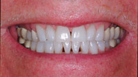
Sometimes a particular case comes along that appears, at first, to be overwhelming. This case fits that description (Figures 1 to 3). However, when this patient e-mailed my office and inquired about the possibility of flying across the country to have me treat him, I had fortunately done many cases involving hundreds of teeth using the matrix system that I developed to treat dentitions afflicted with black triangles, albeit none of this magnitude. I felt absolutely confident that we could achieve a good outcome. The trick was to disassemble the case into bite-sized pieces.
This case presents many excellent questions and the additional challenge of severe facial abrasions. I will first review the background of black triangles and of lower incisor complications and then proceed with the presentation of the clinical procedures used to treat this particular patient.
BLACK TRIANGLES: PREVALENCE AND PATIENT ATTITUDES
One third of adults have unaesthetic black triangles, which are more appropriately referred to as open gingival embrasures.1Besides being unsightly and prematurely aging the smile, black triangles are prone to accumulate food debris and excessive plaque.2 A recent study of patient attitudes found patient dissatisfaction with black triangles to rank quite highly among aesthetic defect, ranking third following carious lesions and dark crown margins.3 If you go online and search "dental black triangles," you will be able to view hundreds of patient black triangle questions and patient complaints/lawsuits resulting from adult orthodontic cases and postperiodontal therapy papilla loss. This clinical and aesthetic dilemma demands more attention from our profession. The caveat is that, until now, there has been no disciplined minimally invasive approach for treatment. Today, instead of improvising and struggling, I have developed a specific predictable protocol to treat this problem.
LOWER INCISOR AESTHETICS
The aesthetics of the lower teeth are often overlooked or simply ignored by many dentists. Recently a fellow passenger seated next to me on a flight was intrigued by the photos that were on my laptop. He asked, "Why do dentists only seem to treat the upper teeth when the lower teeth look all jacked up? Do they think no one notices? It looks ridiculous to have perfect top teeth and ugly bottom teeth!" In addition, as we age, the lower incisors become more visible as the facial muscles lose their tension on the lower lip.
LOWER INCISOR CHALLENGES AND ETHICS
Lower incisors present their own unique restorative challenges. The incisal edge is broad and thin mesiodistally. The root, in contrast, is very broad buccolingually. Imagine a butter knife that has been permanently twisted at 90° in the middle of the blade. This anatomic curiosity creates demanding draw/path of insertion issues for a porcelain laminate or full-crown preparation. A lower incisor with significant recession leads to a mutilatory tooth preparation for porcelain. When I had an opportunity to show this case to the top ceramists in Toronto, Ontario and Seattle, Wash, their answer was refreshingly candid: "Dr. Clark, to treat this case properly with porcelain laminates would require you to mutilate these teeth."
 |
 |
| Figure 1. Preoperative view of a black triangle case. Note the pursing of lips and forced smile of a patient who is embarrassed of the aesthetics of the lower teeth. | Figure 2. The receded papilla height of the anterior teeth was not significantly lower than that of the posterior teeth, ruling out a surgical approach. |
 |
| Figure 3. This view demonstrates the unique "twisted butter knife" anatomy of the lower incisor tooth. |
 |
 |
| Figure 4. High magnification (8x) of the cementoenamel junction area of the tooth. This area is virtually impossible to clean with a prophy cup and scaler, and virtually unbondable unless the dentin is clean and the surface abraded. | Figure 5. High magnification (12x) view of the root after step 9. Note how the gentle blasting has stripped away the contaminated surface dentin and yet leaves the enamel almost undisturbed. |
 |
 |
| Figure 6. Bioclear "Prophy Plus" unit snaps to the quick disconnect, and this or a prophy-jet should be part of every bonded procedure’s armamentarium. | Figure 7. Close-up view of the blasting of the difficult to clean areas. They should also receive the same attention from the lingual aspect (not pictured). |
 |
 |
| Figure 8. Step 9 view at low magnification. Facial surfaces that previously had large abrasions are at full contour. Cord is still in the sulcus but not visible in photograph. | Figure 9. Yellow ContacEZ (Contact EZ) lightens the contact, allowing insertion of the matrix and at the same time removes calculus and plaque from the contact area. So integral to the technique, they are now included in the Bioclear Matrix kit. |
 |
 |
| Figure 10. Bioclear Matrix system complete kit includes diastema closure, anterior, and posterior matrices. Mild to wild emergence profiles are coupled with different tooth sizes and incisal shapes. Sabre wedges, interproximators and other essentials round out the kit. |
Figure 11. A Bioclear DC-202 matrix is ready to be placed incisogingivally once the contact is lightened. Note the curved Incisal edge and aggressive cervical curvature. |
 |
 |
| Figure 12. The DC-203 matrix that is especially designed for diastema closure of small teeth. Side view and profile views are featured. Note the straight incisal edge and the aggressive cervical curvature. |
Figure 13. Four sectional matrices (Bioclear DC-203 matrices) are placed incisogingivally after the contact areas were lightened and gently abraded. |
 |
 |
| Figure 14. A 37% Phosphoric Acid Etchant (3M ESPE) is injected under the matrix on to the tooth. The entire tooth should be etched. | Figure 15. A familiar site to Bioclear users, yet perhaps odd to any "newcomers." The injection molded restoration has interproximal areas that are "porcelainesque" with smooth, rounded contours and flawless surfaces. |