Home › Forums › Pedodontics › AVULSED TOOTH TREATMENT OPTIONS
Welcome Dear Guest
To create a new topic please register on the forums. For help contact : discussdentistry@hotmail.com
- This topic has 6 replies, 2 voices, and was last updated 16/03/2012 at 6:09 pm by
Drsumitra.
-
AuthorPosts
-
25/01/2012 at 5:39 pm #10275
 drmithila
OfflineRegistered On: 14/05/2011Topics: 242Replies: 579Has thanked: 0 timesBeen thanked: 0 times
drmithila
OfflineRegistered On: 14/05/2011Topics: 242Replies: 579Has thanked: 0 timesBeen thanked: 0 timesIt is every general dentist’s nightmare. At 4 o’clock after a busy day, a frantic mother calls telling your receptionist that her 6-year-old son has knocked out his front permanent teeth in a playground accident. When the bloody, crying child arrives at your office, the mother hands you a cup of milk with teeth Nos. 9 and 10 at the bottom. Upon clinical examination, you realize that not only has the child avulsed his central and lateral incisors, but the alveolar bone on both teeth has broken out both buccally and palatally. After numbing the now hysterical child, an attempt to reimplant the teeth seems useless because (1) tooth No. 8 is the only decent tooth to bond to and (2) replacing the teeth into broken sockets feels like placing teeth into Jell-O. Despite your best effort to compress periosteum and alveolar bone around the teeth to stabilize them, the likelihood of successful reimplantation is poor.
Such an occurrence is a long-term personal and family tragedy emotionally, aesthetically, and financially. The probable outcomes are apexification, endodontic treatment, eventual extraction, and a “flipper” partial, bridgework, or implant.
This article will discuss a new approach to stabilizing avulsed teeth that the author believes may have benefits not only for this application, but for other dental trauma situations as well.THE “UGLY DUCKLING” STAGE
The ugly duckling stage is an awkward period in a child’s life when newly erupted maxillary incisors appear to be “snow shovels” in a small face. This is a particularly hazardous time for traumatic dental injuries because of the size and protruding nature of the front teeth, the softness of the alveolar bone, the lack of completed radicular formation, and the lack of ability to splint to adjacent teeth.
Frequently, avulsed teeth occur in young patients with predisposing orthodontic conditions such as thumb-sucking and/or class II malocclusion with excessively flared maxillary central incisors. These patients have maxillary incisors that hang over the lower lip, and when the child falls down, his or her teeth strike first without lip cushion to soften the blow.
I’ve known orthodontists who routinely extract a tooth with a history of avulsion. These teeth frequently ankylose and often fail long term. They feel it is better to correct the problem in the patient’s teens than to retreat the problem in the patient’s 30s or 40s.
When I saw the child described in the opening paragraph, the next day after trying to splint avulsed teeth Nos. 9 and 10 to teeth Nos. 8 and 11, I realized there was no hope. The orthodontic wire and composite were removed from the hanging teeth, and the patient was fitted with an occlusal guide. The appliance was inserted over the loose teeth.25/01/2012 at 5:39 pm #15089 drmithila
OfflineRegistered On: 14/05/2011Topics: 242Replies: 579Has thanked: 0 timesBeen thanked: 0 times
drmithila
OfflineRegistered On: 14/05/2011Topics: 242Replies: 579Has thanked: 0 timesBeen thanked: 0 timesTHE OCCLUSAL GUIDE
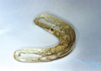
The occlusal guide is a preformed polypropylene orthodontic positioner. For decades, orthodontists have used custom-made positioners to fine-tune orthodontic detail into their fixed orthodontic cases. Orthodontic labs manufacture custom-made positioners by cutting out every tooth from a plaster cast of a nearly completed orthodontic case. The cut-out plaster teeth are reassembled into perfect occlusion in ideal tip, torque, and angulation at Angle class I interdigitation. Then a polypropylene, football-style mouth guard is fabricated over the ideally reset plaster cast.
Dr. Earl Bergersen, an orthodontist and developer of the occlusal guide, realized how extremely uniform the human dentition is. This is why preformed denture teeth can precisely restore ideal aesthetics to an edentulous patient and why aesthetic dentistry has adopted the “golden proportions” in cosmetic makeovers. Prefabricating the positioner by duplicating different sizes of denture teeth set to ideal class I occlusion eliminated the need for orthodontists to remove the last upper and lower wires, take an impression, replace the same wires, send the poured models to the lab for positioner fabrication, and have the patient return for debonding, debracketing, and appliance insertion. Orthodontists could now select the size needed and insert the best-fitting appliance immediately after fixed appliance removal. The patient needs to do heavy biting exercises for a prescribed length of time, say 2 hours per day for one week, prior to final retention.
Dr. Bergersen soon realized that his device with a motivated child could do far more than provide fine detail to a near-finished orthodontic case. He found that the occlusal guide was capable of correcting severe orthodontic malocclusions with sustained use in a motivated child. Huge overjet/overbite cases could be corrected 1 mm per month. Severely crooked teeth with absence of crowding could be straightened over several months of heavy biting exercises. The flexible but firm polypropylene rims can catch misaligned teeth and slowly force them into place. The occlusal guide became the most frequently used orthodontic appliance worldwide.THE AVULSED TOOTH STABILIZING APPLIANCE
The problem with using an occlusal guide to stabilize avulsed teeth is that it is both an upper and lower bulky, football-style rubber mold. The patient has to eat and drink, necessitating early removal during the crucial 7- to 10-day period for avulsed teeth. Early removal could possibly remove the teeth with the appliance, because dried blood and contaminants cake to the very loose teeth, and the appliance slots grasp tooth curvatures, making removal risky.
However, if the mandibular half was eliminated from the appliance, leaving only the upper, then with some difficulty the patient could eat and drink for days, perhaps weeks, before removing the occlusal guide (see Figure). Longer-term wear may allow complete reattachment of the avulsed teeth. Intermittent biting exercises would place an apically directed force, maintaining the terminal root in the depth of socket position at an ideal tip, torque, and angulation. I estimate that 3 sizes of the appliance would fit 90% of patients: small, medium, and large. The size is determined by arch width and central incisor width.
Frequently, pediatric patients age 6 to 8, in their ugly duckling stage, have teeth ectopically positioned with flared centrals and huge diastemas. Because of the very flexible material used to fabricate the occlusal guide, these teeth can still be stabilized by the appliance even if the central incisors overlap into the lateral slots. Again, the occlusal guide was designed to straighten crooked teeth. Different size appliances may be inserted to determine the best fit, although this appliance will not fit all mouths.The patient should be instructed to wear the appliance continuously for 7 to 10 days including eating, drinking, and sleeping, while intermittently biting hard into the appliance throughout the waking hours as much as possible. Sleep could be difficult, especially the first night, and as with any oral appliance, temporary excessive salivation is a frequent problem.
After 7 to 10 days, the child should be re-examined. Then the dentist should peel back the phlanges of the appliance labially and palatally, detaching any dried blood and contaminants. The teeth should be examined for stability and firmness. If after 7 to 10 days a firm reattachment is not evident, the teeth should be removed.
Besides being useful for tooth reimplantation in the ugly duckling stage, this device could be used for multiple avulsed teeth that are difficult to stabilize. For example, I had a patient who avulsed all 4 maxillary incisors when his teeth caught the net while dunking a basketball. The attempt to reimplant and stabilize them by an oral surgeon was unsuccessful. Stabilizing multiple teeth avulsions in a bloody operating field with composite and orthodontic wire can be very difficult. The patient now has 4 implants.
This device could also remedy other traumatic dental injuries, such as accident victims where the incisors are crushed palatally or are very loose but not fractured. These teeth could be repositioned to close to an ideal position and the appliance inserted for 7 to 10 days.25/01/2012 at 5:40 pm #15090 drmithila
OfflineRegistered On: 14/05/2011Topics: 242Replies: 579Has thanked: 0 timesBeen thanked: 0 times
drmithila
OfflineRegistered On: 14/05/2011Topics: 242Replies: 579Has thanked: 0 timesBeen thanked: 0 timesPerhaps this device could be useful for fractured teeth; broken teeth stabilized precisely may allow successful root canal treatment, especially with current perforation sealing techniques. I propose the hypothesis that sustained, apically directed pressure on the crown into the bony socket of a mid root fracture would more likely correctly align a fractured root than a dentist bonding a loose crown haphazardly to adjacent teeth with orthodontic wire.
DISCUSSION
Many gaps exist in current treatment for successful reimplantation of avulsed teeth, even if the accident victim arrives to a medical professional within the narrow window of time needed. For example, general dentists see these cases so infrequently that they are often ill-suited to treat a screaming, bloody, ugly duckling child with no adjacent teeth to which to bond. They often refer them to an oral surgeon across town.
In car accidents with multiple avulsed teeth, medical per-sonnel are rarely prepared to handle anything dental. Even large trauma centers with around-the-clock general surgery, orthopedic, and cardiovascular residency coverage often call in oral surgeons to handle tooth avulsions. What if every paramedic had 3 sizes of the avulsed tooth stabilizing splint on board his vehicle and was trained to reimplant and insert the appliance even as a temporary means to stabilize? What if every emergency room physician/resident or general dentist had these appliances on hand ready to use?
Perhaps unconscious accident victims could be stabilized by reimplanting the teeth, selecting and fitting the appropriate appliance, relining the splint with cold care liquid gel polypropelene, and reinserting the appliance over avulsed teeth until the material was hardened and secure.CONCLUSION
How many avulsed teeth are lost by holes in the healthcare system? How many avulsed teeth are lost by poor stabilization of multiple avulsed teeth with little to which to bond? How will prolonged, intermittent, apically directed biting force affect reimplantation success in the short term and long term? Can nondental, first-arriving healthcare professionals be trained to reimplant avulsed teeth with a preformed polypropylene mold? Will mid root fractures have a greater chance of success if the broken fragments are compressed over each other for 7 to 10 days? Will this device help other dental injuries such as subluxation and tooth loosening?
The answers to these questions won’t be known for perhaps years. However, how many thousands of avulsed teeth are failing currently? Failure to reimplant an avulsed tooth correctly can doom a young child and the family to a long emotional, financial, and aesthetic tragedy. This article has discussed a new treatment ap-proach that the author feels has promise for stabilizing avulsed teeth and for certain other dental trauma situations as well. Additional clinical experience is needed to determine how successful this approach will be.25/01/2012 at 5:40 pm #15091 drmithila
OfflineRegistered On: 14/05/2011Topics: 242Replies: 579Has thanked: 0 timesBeen thanked: 0 times16/03/2012 at 6:08 pm #15286
drmithila
OfflineRegistered On: 14/05/2011Topics: 242Replies: 579Has thanked: 0 timesBeen thanked: 0 times16/03/2012 at 6:08 pm #15286Drsumitra
OfflineRegistered On: 06/10/2011Topics: 238Replies: 542Has thanked: 0 timesBeen thanked: 0 timesDenture stabilization with implants can make a dramatic difference in the lives of patients, providing benefits in function, aesthetics, and overall health. However, for many denture wearers, traditional implant treatment may be unattainable for any number of reasons. A primary factor is frequently the expense of the procedure. Inadequate bone can be a challenge for many patients, requiring extensive bone grafting prior to conventional implant placement. Finally, as patients age, many simply do not wish to devote a great deal of time to a surgical process that can go on for months and requires a considerable amount of recovery time.
As a general practitioner who has an extensive history placing conventional dental implants, I am an enthusiastic advocate for the traditional procedure. However, I have seen many patients in my practice for whom it is impractical or simply out of reach financially. The last few years have seen a trend developing for a different kind of implant treatment that may provide an excellent solution for some denture patients involving mini-dental implants (MDIs) (also known as small-diameter implants).
MDIs were initially introduced as transitional devices to retain a denture while a conventional implant was allowed to osseointegrate. What many practitioners found was that if a patient did not return to have these transitional implants removed within 3 to 6 months, they became very difficult to remove, as they too had integrated into the bone.1 In 2004, the FDA-approved MDI System (3M ESPE) (formerly IMTEC Sendax MDI implants) for long-term use.
In recent years, this treatment has been increasingly discussed by the implantology community, primarily as a solution for patients who are not ideal candidates for conventional implants or who cannot afford this option. In my practice, it has been particularly appropriate for patients with atrophic mandibles who do not wish to go through the expense or time of conventional implant treatment with significant bone grafting.
The official protocol for placing MDIs is taught to general practitioners as well as specialists in one-day seminars, making it a relatively simple technique to learn. A minimum of 4 implants are recommended for mandibular denture stabilization. The sites for each implant are marked on the patient’s tissue, and a 1.1-mm pilot drill is used to create entry points. The mini-implants are inserted into the pilot holes and then advanced with a progression of a finger driver, winged thumb wrench, and a ratchet. As a clinician with significant experience in implant placement, I use a more advanced procedure utilizing a flap in cases if appropriate, but a basic case can typically be performed without this step. After placement of the implants, the patient’s denture is then fitted with housings that snap onto the o-ring heads of the implants. This allows the denture to be tissue supported but implant retained, which offers the capability of immediate loading.
Reported success rates for MDIs have ranged from 91% to 97.4%.2-5 The most comprehensive study tracked 2,500 implants and reported a 5-year survival rate of 94.2%.4 As the body of research for this treatment grows larger, additional evidence can be expected to support the suitability of MDIs in the edentulous mandible16/03/2012 at 6:09 pm #15287Drsumitra
OfflineRegistered On: 06/10/2011Topics: 238Replies: 542Has thanked: 0 timesBeen thanked: 0 times

Figure 7. MDIs impression caps were luted with light-cured flowable resin (Heliomolar [Ivoclar Vivadent]). Figure 8. The maxillary denture and mandibular implant overdenture setup (in wax) (Blueline Teeth [Ivoclar Vivadent]). 

Figure 9. The implants, 4 weeks postsurgical. Figure 10. Mandibular overdenture with housings for the o-rings in the undersurface. 

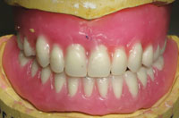
Figure 11. Final panoramic radiograph. Figure 12. Completed dentures in centric occlusion. 16/03/2012 at 6:09 pm #15288Drsumitra
OfflineRegistered On: 06/10/2011Topics: 238Replies: 542Has thanked: 0 timesBeen thanked: 0 timesPostdelivery Appointments
Follow-up appointments have shown the patient to be thrilled with the treatment. She stated that it had made a huge change in her life and had given her much more confidence. After experiencing the level of stability made possible with the MDIs in the mandible, the patient is now considering a similar procedure for the maxilla. Despite the fact that stability in the maxilla was not an initial concern for the patient, she now feels that if it can be made better, she would like to pursue treatment to improve her confidence even more.
DISCUSSION
This case demonstrates 2 variances from the standard protocol for MDI placement, in that a flap was performed and the implants were not immediately loaded. As a dentist who has been traditionally trained in implant placement, I personally prefer to create flaps in cases with atrophic mandibles. While not strictly required for MDI placement, a flap allows the clinician greater certainty of placement in the middle of the crest. In cases where more bone is available, a flapless procedure is quite straightforward.
Because the patient in this case was relatively young, the decision was made to not immediately load the implants in order to allow the bone and soft tissue to mature more fully. This simply provides more assurance that the implant will survive in the long-term with a young patient in robust health. Immediate loading is often very suitable for older patients, due to the fact that their occlusal forces may not be as strong, and they are seeking an immediate quality of life improvement rather than an implant that will survive for 10 years or more. However, in this case it was determined to allow for a longer period of bone maturation prior to engaging the retentive feature of the overdenture. Fixation of the implant at placement is an essential requirement for success of the MDI system, as well as with conventional endosseous implants.
It is critical that the clinician utilize an array of different clinical findings and technology to assist with long-term treatment decisions. Most recently, I have incorporated the usage of the Periotest (Medizintechnik Gulden). While not required in the MDI protocol, I am using it in addition to a torque wrench to establish another quantitative value prior to immediate load cases, as it provides additional information. It is also a test that can be performed throughout the life of the implant, which helps me follow implant specific integration over time. Most importantly, however, is that the implant after placement demonstrates zero mobility visually upon percussion.
In this case report, a moderate divergence of the left implants is exhibited in the final panoramic radiograph. This clinical result occurred despite parallel 1.1-mm drills placed in the osteotomies prior to implant placement. This clinical finding can occur due to several reasons, including the partial osteotomy protocol, self-tapping nature of the implant, quality of bone, and the clinician’s surgical decision making. The partial osteotomy surgical protocol combined with the self-tapping nature of the implant and soft bone can allow for minor variations in the implant path. It is essential for the novice or experienced clinician to guide the placement of the implant in the path desired for an ideal outcome. The presence of anatomical structures such as the mental foramen and a potential anterior loop of the inferior alveolar nerve may dictate implant placement. Therefore, it is very common to see a distal implant divergence, due to the clinician’s tendency to position the implant mesial to the neurovascular complex. Finally, divergence of implants is successfully managed by the versatility of the MDI system’s MH-1 o-ring housing design. This prosthetic attachment design allows for a firm retentive feature within a 30° implant divergence. The patient has been seen on a 4-month recall basis for the past 2 years, demonstrating excellent retention, minimal o-ring wear and excellent crestal bone levels. Most importantly, the patient feels that the implant-retained overdenture is a huge success.CONCLUSION
Many dentists have likely seen denture patients who have suffered great losses in their quality of life, and have been making do with temporary measures like adhesives and over-the-counter relines for far too long. MDIs give dentists an important tool to reach this pool of patients and provide them with an affordable and less invasive path to denture stabilization. As the patient in this case demonstrates, added stability can bring back the quality of life to a large population of patients. -
AuthorPosts
- You must be logged in to reply to this topic.
