Home › Forums › Oral & Maxillofacial surgery › EXTRA-ORAL EXAMINATION FOR CANCER DETECTION
Welcome Dear Guest
To create a new topic please register on the forums. For help contact : discussdentistry@hotmail.com
- This topic has 3 replies, 2 voices, and was last updated 03/12/2012 at 5:23 pm by
 drmithila.
-
AuthorPosts
-
30/12/2011 at 1:51 pm #10241
Drsumitra
OfflineRegistered On: 06/10/2011Topics: 238Replies: 542Has thanked: 0 timesBeen thanked: 0 timesEXTRAORAL AND INTRAORAL EXAMINATIONS
The following is a concise overview of the components of the extraoral and intraoral examination. It stresses a systematic and consistent approach to these examinations. (The order of the examination steps as described herein, is the systematic sequencing that the author uses. The order of the examination steps may vary depending individual clinician-determined protocols.)
Systematic Extraoral Examination
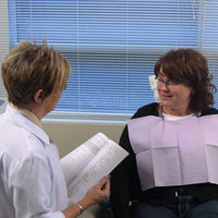
A review and assessment of the systemic health and pharmacological status of the patient is always done prior to any dental examination. The extraoral examination continues with observation of the head and neck, as well as observation of the sound of the patient’s voice and eye movements commencing from when the patient is first seated in the treatment room (Figure 1). Hoarseness in the voice may warrant further investigation if it has been persistent, since this may be an indication/suspicion of a growth within the larynx/oropharynx. Abnormal breathing may be a sign of anxiety or fatigue. Pupil size may signify a reaction to drugs or state of emergency as well as an indication of a disease state or inflammatory presence. The appearance of the face is further evaluated noting any asymmetry, swelling or discoloration. Inspection of the skin includes the color, texture, the presence of eruptions or swellings, or any abnormal growth. Observe all areas of exposed skin, paying particular attention to areas behind the ears and the back of the head and neck. Most people will have freckles, birthmarks, or moles; irregularities or a change in the shape, edge, color, and/or size can be a warning sign of skin cancer thus warranting further investigation.
Have your patients remove their eyeglasses to make certain there are no hidden growths or developments that would have otherwise gone unnoticed. The areas along the hairline and under the eyeglasses will require tactile palpation in order to discern or identify any swellings/growths.

Figure 1. Initial observation of head and neck, speech, and eye movements. Figure 2. Examination of the temporomandibular joint. 

Figure 3. Bilateral palpation of parotid
salivary glands.Figure 4. Bilateral palpation of submental nodes. 

Figure 5. Bilateral palpation of submandibular nodes. Figure 6. Bilateral palpation of cervical lymph nodes. 

Figure 7. Bilateral palpation of supraclavicular nodes. Figure 8. Bilateral palpation of occipital nodes. 

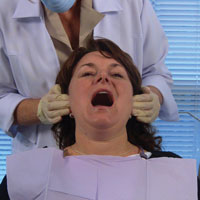
Figure 9. Bilateral palpation of postauricular nodes. Figure 10. Bilateral palpation of preauricular nodes. Next is the examination of the temporomandibular joint, utilizing a bilateral examination technique (Figure 2). This is accomplished by placing your finger pads over the joint just anterior to the ear; instructing the patient to open and close as well as move the jaw to the left and right; checking for any limitations or deviations upon opening, subluxation, any tenderness, sensitivity or any noises such as a grating, clicking, or popping.
The next area to be examined is the parotid salivary glands (Figure 3). The extraoral palpation of the parotid salivary glands is best examined using a bilateral technique, employing light pressure and placing fingers at the angles of the mandible over the parotid glands. Compare the bilateral findings for symmetry. Normal parotid glands are not palpable and exhibit no tenderness. Abnormal salivary glands may be painful, swollen, and indurated.
The lymph nodes are examined next with the clinician behind the patient and the patient’s chin slightly elevated. Areas of particular concern in a systematic examination can be found in the Table.
It is important to inform the patient as to the relevance of the examination of the lymphatics of the head and neck before commencing this portion of the extraoral examination. In addition, one should indicate what areas of the head and neck will be examined. Due to the diverse multiculturalism that exists within our patient population, we must be culturally aware and sensitive to the different possible comfort levels of our patients.
Evaluation of the lymph nodes is done by a gentle rolling motion of the fingers, using the bilateral palpation technique. Note any enlargement, tenderness, lack of mobility, hardness, or asymmetry. If enlargement is detected, the examiner should determine the mobility and consistency of the nodes. Enlargement or lymphadenopathy may be attributed to either an infectious or inflammatory process or a malignant neoplasm. Clinical characteristics can help discern the difference.
In the broadest clinical terms, the enlarged node, if related to infection, is most often soft, freely movable, and painful. Also, the patient may have presented with an infection (or presence of inflammation) and may occasionally possess some knowledge of the etiology. Malignant neoplasm related nodes are normally fixed, particularly in the later stages, and they are generally not painful. One could compare the consistency of an infection related node to a blueberry or pea, whereas a malignant neoplasm related node is normally firmer in consistency, like a stone.
Next, submental and submandibular nodes should be examined carefully. With the patient’s head back slightly, first examine the submental nodes (Figure 4). Instruct the patient to bite together lightly and place the tongue into palatal vault. This results in a tensing of the mylohyoid muscle, allowing for easier palpation of submental glands. Moving posterior toward the angle of the mandible and palpating directly below the line of the mandible are the submandibular glands (Figure 5).
Another area to examine are the cervical nodes; both superficial and deep nodes. This set forms a complex chain of numerous nodes. Instruct the patient to turn the head in order to reposition the sternocleidomastoid muscle for ease of palpation and better access of both the superficial/deep cervical nodes (Figure 6).
The supraclavicular nodes are palpated next, found superior to the clavicle in the hollow area or supraclavicular fossa directly above the collarbone (Figure 7). They drain a part of the thoracic cavity and abdomen. Virchow’s node is a left supraclavicular node, which receives the lymph drainage from most of the body (especially above the abdomen) via the thoracic duct; this node may serve as an early site of metastasis for various malignancies.
The next nodes to be palpated are the occipital nodes (Figure 8). These are associated with the occipital artery at the posterior base of the skull. Using a bilateral technique, palpation is done directly below the base of the occipital bone. Reclining the patient’s head to the front, exposing the occipital area may facilitate better access for palpation of the occipital nodes.
The posterior auricular, or postauricular, nodes are next in the systematic order of lymph node palpation and are usually 2 in number (Figure 9). The anterior auricular or preauricular nodes are from one to 3 in number and lie immediately in front of the tragus (Figure 10). Both pre- and postauricular nodes’ efferent vessels drain into the superior deep cervical nodes.
The thyroid gland, normally not detected by palpation, is examined next.
An abnormal gland could be indurated, enlarged on one or both sides, or contain palpable nodes. When using bilateral palpation, palpation is done on both sides of the gland, noting any nodules or masses (Figure 11). Instruct your patient to swallow, which in turn will elevate the thyroid gland; allowing for an abnormality to become more apparent. Asymmetrical movement of the thyroid cartilage during swallowing might indicate that the gland is fixed to underlying tissues. If the patient is obese, it may be easier to palpate this area positioned behind the patient, having him or her turn the head toward the examining side. Suspicious thyroid gland findings should be referred to your patient’s physician for further evaluation.09/08/2012 at 5:41 pm #15801Drsumitra
OfflineRegistered On: 06/10/2011Topics: 238Replies: 542Has thanked: 0 timesBeen thanked: 0 timesNew analysis suggests that for some people with high risk factors, basal cell carcinoma is a chronic disease.
High sun exposure before the age of 30 was a major predictor, as was a history of eczema.
“Basal cell carcinoma is a chronic disease once people have had multiple instances of it, because they are always at risk of getting more,” says Martin Weinstock, professor of dermatology in the Warren Alpert Medical School of Brown University, who practices at the Providence Veterans Affairs Medical Center.
“It’s not something at the moment we can cure. It’s something that we need to monitor continually so that when these cancers crop up we can minimize the damage.”
Dermatologists hold out hope for a medication that will help prevent recurrences of BCC. To test one such medicine, Weinstock chaired the six-site, six-year VA Topical Tretinoin Chemoprevential Trial, which last year found that the skin medication failed to prevent further instances of BCC in high-risk patients.
10/10/2012 at 5:40 pm #16009 drmithila
OfflineRegistered On: 14/05/2011Topics: 242Replies: 579Has thanked: 0 timesBeen thanked: 0 times
drmithila
OfflineRegistered On: 14/05/2011Topics: 242Replies: 579Has thanked: 0 timesBeen thanked: 0 timesA new gene test may end up saving many lives.
The new test can detect precancerous cells in patients with benign-looking mouth lesions. This test may open the possibility for at-risk patients to receive treatment earlier than they would have before this test was created. The result would be a greater chance of survival.
The study appeared online in the International Journal of Cancer. The results indicated that the test was correct at a rate of 91 to 94 percent of the time. There were 350 head and neck specimens studied.
The amount of people who have mouth cancer is about 500,000 worldwide and the number could reach one million by 2030, according to some studies.
Only about five to 30 percent of mouth lesions will develop into cancer. The cancer can be treated if detected early enough. There hasn’t been a test that can accurately detect which lesions will become cancerous at this point.
The current diagnosis method is histopathology, which tests biopsy tissue taken during an operation. A pathologist analyzes the tissue under a microscope. Because of the invasive nature of the procedure, most mouth cancers were diagnosed at later stages.
More clinical trials are necessary to determine long-term benefits of this test. It’s conceivable that this test will eventually be used to detect types of cancer other than mouth cancer.
03/12/2012 at 5:23 pm #16221 drmithila
OfflineRegistered On: 14/05/2011Topics: 242Replies: 579Has thanked: 0 timesBeen thanked: 0 times
drmithila
OfflineRegistered On: 14/05/2011Topics: 242Replies: 579Has thanked: 0 timesBeen thanked: 0 timesThe genesis for the test came from a collaboration between Neil Gottehrer, DDS, a Havertown, PA, dentist who has been practicing for 38 years, and David T.W. Wong, DMD, DMSc, a pioneer in salivary diagnostics who is the associate dean of research and a professor of oral biology at the University of California, Los Angeles (UCLA) School of Dentistry.
Neil Gottehrer, DDS, PeriRx co-founder.
Dr. Wong’s lab has been looking at salivary diagnostics for a range of illnesses, including pancreatic and lung cancers and type 2 diabetes, and he is involved in a multidisciplinary effort to create a point-of-care device for analyzing the biological markers of disease in saliva.“I saw what he was able to do with oral cancer markers and what an impact it had,” Dr. Gottehrer told DrBicuspid.com. “As a dentist, we’ve been trained to do cosmetic dentistry, and we would now have an opportunity to save lives.”
Dr. Gottehrer then discussed the idea of a saliva test for oral cancer with his patients, Jack Martin, MD, a cardiologist from Bryn Mawr, PA, and Stephen Swanick, who became company CEO.
“They were pretty one-sided conversations,” Dr. Martin joked of the chairside exchanges, which took place while Dr. Gottehrer worked on his teeth. Together, the two founded PeriRx in 2008.
Drs. Gottehrer and Martin had worked on other projects together, including one that investigated whole-body inflammatory connections.
“It’s much better to create a company with patients you treat and respect professionally, so this was a natural for us to work together,” Dr. Gottehrer explained.
Specific to OSCC
The molecular marker test differs from fluorescence-based imaging technologies and other testing devices because it provides an objective quantification with no subjective decisions by dentists, Dr. Martin said.
“The other tests require a lot of judgment by the practitioner about the color of lesions,” he told DrBicuspid.com. “Our test produces a quantitative result with test scores that helps decide who needs to have a lesion biopsied right away because of the high chance of malignancy versus lesions that just need to be watched. So you’re not doing a lot of unnecessary biopsies on people who don’t need it.”
Head and neck squamous cell cancer is the sixth most common type of cancer worldwide, and almost 600,000 cases are reported annually; of these, approximately 10% are oropharyngeal squamous cell carcinoma (OSCC), according to the U.S. Centers for Disease Control and Prevention.
Nearly 40,000 Americans are diagnosed with oral cancer annually, according to the Oral Cancer Foundation, and more than 8,000 die each year from the disease. Early detection of OSCC can result in 90% survival rates, but most U.S. cases are not diagnosed until stage 3, when the five-year survival rate plunges to about 43%, Swanick noted.
“As a dentist, we’ve been trained to do cosmetic dentistry, and we would now have an opportunity to save lives.”
— Neil Gottehrer, DDS
About 35% of dentists screen patients for oral cancer, starting with a visual exam for lesions and often followed by fluorescence-based imaging technologies, according to Swanick.A handful of different testing devices are on the market, but none has improved the survival rate, he said. “That’s very, very daunting,” Swanick said. “What we’re really chasing here is a 40% improvement in the survival rate that will equate into saving more lives.”
Other saliva tests check for the presence of HIV and human papillomavirus (HPV), but the PeriRx test is designed specifically to identify OSCC, he added, noting that about 90% of oral cancers are squamous cell carcinoma.
Dentists are often at the vanguard for detecting oral cancer because most patients see them more frequently than their primary care doctors, Dr. Martin noted.
“I have patients I see [for heart problems] who tell me they haven’t seen their family doctor in 10 years, but they see their dentist every four months,” he said. “That’s why dentists are becoming more of a primary healthcare provider in screening patients for a number of systemic diseases.”
With PeriRx, hygienists would collect about 3 cc of saliva and send it for lab analysis, which will also check for periodontal disease. Results would be available within 48 hours, according to the company.
“This brings hygienists much more closely into patient-involved treatment because they’ll take the test and discuss the potential impact on their overall health,” Dr. Gottehrer said, noting that several hygiene program directors have described this as hygienists becoming dental nurses.
“Hygienists are very anxious to participate,” he said. “And it will take the responsibility off the dentist, but he makes the final decision, so it’s a team effort.”
Insurance companies are interested in seeing this type of technology since early detection of oral cancer reduces costs across the board, said Dr. Gottehrer, adding that many insurers now cover saliva tests.
Clinical testing
The PeriRx test was evaluated by studies that included more than 500 patients at UCLA and also patients in Serbia and India, according to data the company provided in response to queries from the National Institute for Health Research (NIHR) in the U.K. The NIHR lists promising new technology that may have a significant impact on British health.
Saliva samples from approximately 300 patients were independently validated by an independent lab of the U.S. National Cancer Institute, the company said.
The PeriRx test has been approved for sale in the European Union. The company is now enrolling patients for clinical trials at Michigan State University and the University of Michigan; the trials are expected to be completed next September. A final assay will be done by the end of 2013, and the company is planning for a U.S. launch in January 2014, Swanick said.
PeriRx plans to pursue a dual regulatory pathway to expedite getting the test to the U.S. market. It will simultaneously apply for laboratory certification and 510(k) premarket submission to the U.S. Food and Drug Administration. The company is in discussions with several large pharmaceutical companies — including GlaxoSmithKline and Johnson & Johnson — about teaming up to distribute the diagnostic test.
PeriRx is also in the process of drafting and creating study protocols for clinical trials testing for type 2 diabetes and prediabetes markers, its second application for the technology.
As more dentists include screening for oral cancer as part of routine exams, they are playing an increasingly pivotal role in finding the disease, Swanick said.
“We applaud the effort of U.S. dentists in the last 15 years to do something to detect this disease,” he said, “And that’s why we think our test will be adopted.”
-
AuthorPosts
- You must be logged in to reply to this topic.