Home › Forums › Endodontics & conservative dentistry › Influence Of White And Gray Endodontic Posts On Color Change
Welcome Dear Guest
To create a new topic please register on the forums. For help contact : discussdentistry@hotmail.com
- This topic has 12 replies, 6 voices, and was last updated 06/01/2012 at 1:15 pm by
Anonymous.
-
AuthorPosts
-
22/02/2010 at 3:59 am #8889
Anonymous
OnlineTopics: 0Replies: 1151Has thanked: 0 timesBeen thanked: 2 timesInfluence Of White And Gray Endodontic Posts On Color Changes Of Tooth Roots, Composite Cores, And All-Ceramic Crowns
Objective: To evaluate whether post materials affect the color of roots, composite cores, and all-ceramic crowns. Method and Materials: Forty extracted human incisors were divided into four groups.
White posts made of zirconia (Zi) or glass fiber (Gf) and gray posts made of titanium (Ti) or carbon fiber (Cf) were randomly assigned to the roots. Composite cores and glass-ceramic crowns were made. The color of the roots, cores, and crowns was captured (Spectroshade).
The mean color difference (mDE) among the groups was calculated for the following comparisons: A—root: empty root versus post and core; B—root: post and core with and without cement; C—core: white versus gray posts and cores; D—lower third of crown versus original ceramic ingot; E—center of crown versus ingot. Statistical analysis was performed by ANOVA, Kruskal-Wallis, and Sheffé tests.Results: White, as well as gray posts, induced little changes of the root color (A, B). Gray posts led to a significant discoloration of the cores (C: mDEZi 2.0 ± 0.7, mDEGf 1.5 ± 0.6, mDETi 12.9 ± 5.9, mDECf 11.2 ± 5.3; P < .0001, Kruskal-Wallis) resulting in a grayish discoloration of the crowns’ lower thirds (D: mDEZi 5.7 ± 0.8, mDEGf 6.0 ± 1.2, mDETi 3.5 ± 1.1, mDECf 3.9 ± 0.9; P < .0001, Kruskal-Wallis). In the center of the crowns, all posts and cores induced a similar color difference (E).
Conclusion: A grayish gingival shadowing cannot be reduced with white posts. In combination with glass-ceramic crowns, white posts and cores are esthetically beneficial.
22/02/2010 at 4:23 pm #13659sushantpatel_doc
OfflineRegistered On: 30/11/2009Topics: 510Replies: 666Has thanked: 0 timesBeen thanked: 0 times22/02/2010 at 7:53 pm #13660Anonymous
here’s something i got from the dentatus website ., ., awaiting more insight from someone who’s been using the luminex posts ., .,
All too often, fragile, thin-walled teeth present major restorative problems: cast posts or extractions were often the only alternative. But today, there is a user friendly, single office visit solution to this problem.
The clear light transmitting posts polymerize light-cured composites within the entire root canal. After curing, the LUMINEX post is removed, leaving a ready canal for a corresponding Classic Post.
* Reinforced Root Strength:
Light-cured composites internally reinforce the root structure providing maximum sheer load support and retention.* Improved Control:
Light-curing composites are easy to control, more adaptive, and safer than auto-cured composites that may prematurely harden.* Centered Canal Position:
The Luminex post technique centers the canal and forms a selected sized, full length parallel sided canal for corresponding Dentatus Classic metal posts.* Superior Aesthetics:
The light-cured composite inside the canal masks metal posts with a reflective tooth colored foundation for modern restorations.* Technique Versatility
Luminex smooth and grooved posts may be also used as an impression and castable post pattern in the direct and indirect fabrication of posts.* Superior Delivery System
Selection of Luminex and metal posts in all sizes along with corresponding reamers and components are packaged in the refillable, easy to use dispenser,01/12/2011 at 4:30 pm #14908 drmithila
OfflineRegistered On: 14/05/2011Topics: 242Replies: 579Has thanked: 0 timesBeen thanked: 0 times
drmithila
OfflineRegistered On: 14/05/2011Topics: 242Replies: 579Has thanked: 0 timesBeen thanked: 0 timesRelyX Fiber Posts offer a unique resin composition and equally dispersed parallel glass fibers, providing superior mechanical strength and dentin-like flex. Its microporous surface allows for additional bond strength through microretentive effects. Its radiopacity is among the highest of available glass fiber posts. With high translucency and an appearance similar to that of tooth structure, RelyX Fiber Posts are not visible through the restoration and support a natural light reflection. The posts are now offered in coronal diameters of 1.1, 1.3, 1.6, and 1.9 mm, providing a solution for any clinical situation. Pairing RelyX Fiber Post with RelyX Unicem 2 Automix Self-Adhesive Resin Cement and its endo tip creates the perfect treatment in endo, enabling cement application from bottom to top. The result is a strong and virtually void-free post cementation.
04/01/2012 at 5:04 pm #15030Drsumitra
OfflineRegistered On: 06/10/2011Topics: 238Replies: 542Has thanked: 0 timesBeen thanked: 0 timesBRIEF HISTORICAL BACKGROUND
For more than 250 years, clinicians have written about the placement of posts in the roots of teeth to retain restorations.1 As early as 1728, Pierre Fauchard described the use of “tenons,” which were metal posts screwed into the roots of teeth to retain bridges.1 In the mid-1800s, wood replaced metal as the post material, and the “pivot crown,” a wooden post fitted to an artificial crown and to the canal of the root, was popular among dentists.1 Often, these wooden posts would absorb fluids and expand, frequently causing root fractures.2 In the late 19th century, the “Richmond crown,” a single-piece post-retained crown with a porcelain facing, was engineered to function as a bridge retainer.2 During the 1930s, the custom cast post-and-core was developed to replace the one-piece post crowns. This procedure required casting a post-and-core as a separate component from the crown.2 This 2-step technique improved marginal adaptation and allowed for a variation in the path of insertion of the crown.1
CAUSES OF POST-RETAINED CROWN FAILURE
The failure of post-retained crowns has been documented in several clinical studies (Figure 1).3 Many of these studies indicate that the failure rate of restorations on pulpless teeth with post-and-cores is higher than that for restorations of vital teeth.3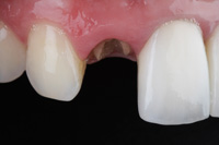
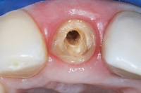
Figure 1a and 1b. Restorative failure of an all-ceramic crown on the maxillary right central occurring after endodontic treatment. A minimum of a 1 mm collar on sound tooth structure is required for a ferrule design.
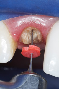
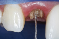
Figures 2a and 2b. After determining the desired post channel length (one half to two thirds length of canal), the gutta-percha was removed with a series of pre-shaping instruments (Gates Glidden [SybronEndo]) (Rebilda post reamer [VOCO]). Several main causes of failure of post-retained restorations have been identified, including: recurrent caries, endodontic failure, periodontal disease, post dislodgement, cement failure, post-core separation, crown-core separation, loss of post retention, core fracture, loss of crown retention, post distortion, post fracture, tooth fracture, and root fracture.4-6 Also, corrosion of metallic posts has been proposed as a cause of root fracture.7
A COMPARISON OF CURRENT POST SYSTEMS
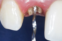
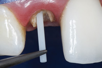
Figure 3. The channel preparation for a prefabricated fiber-reinforced post was performed using a color-coded drill (Rebilda post drill [VOCO]), establishing the desired intraradicular length and size for the selected post.
Figure 4. The pre-selected fiber-reinforced composite post (Rebilda post [VOCO]) was placed into the channel space. The coronal height was measured and marked with a diamond disc to the desired length. The post is cleaned with alcohol, silanated (Ceramic Primer [VOCO]) for 60 seconds, and then air-dried.
Today, the clinician can choose from a variety of post-and-core systems for different endodontic and restorative requirements. These systems and methods are well-documented in the literature.8-10 However, no single system provides the perfect restorative solution for every clinical circumstance, and each situation requires an individual evaluation.
Custom Cast Posts
The traditional custom-cast dowel core provides a better geometric adaptation to excessively flared or elliptical canals, and almost always requires minimum tooth structure removal.1 Custom cast post-and-cores adapt well to canals with extremely tapered canals or those with a noncircular cross section and/or irregular shape, and roots with minimal remaining coronal tooth structure.9 Patterns for custom cast posts can be formed either directly in the mouth or indirectly in the laboratory. Regardless, this method requires 2 appointment visits and a laboratory fee.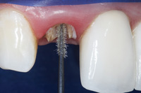
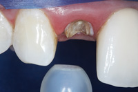
Figures 5a and 5b. A dual-curing, self-etch adhesive (Futurabond DC [VOCO]) was applied with an applicator (Endo Tim [VOCO]) to the base of the post space and air-dried. Any excess adhesive was absorbed with an endodontic paper point using a rapid
intermittent movement.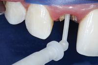
Figure 6. A dual-cure, resin cement (Bifix QM [VOCO]) was injected into the post channel using an angled tip (Intraoral Tip Type 1 [VOCO]). It is important to remove the tip slowly while injecting, to prevent incorporation of air bubbles.
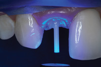
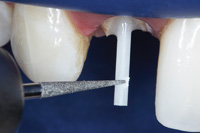
Figures 7a and 7b. The fiber post was immediately inserted into the post hole to the base of the prepared channel and light-cured from different positions for 2 minutes (7a). After polymerization, the fiber post was cut with a diamond bur to the predetermined length. Never use a serrated instrument or shears because this can damage the integrity of the post(7b). Also, because it is cast in an alloy with a modulus of elasticity that can be as high as 10 times greater than natural dentin,11 this possible incompatibility can create stress concentrations in the less rigid root, resulting in post separation and failure. Additionally, the transmission of occlusal forces through the metal core can focus stresses at specific regions of the root, causing root fracture.11Furthermore, upon aesthetic consideration, the cast metallic post can result in discoloration and shadowing of the gingiva and the cervical aspect of the tooth.
04/01/2012 at 5:05 pm #15031Drsumitra
OfflineRegistered On: 06/10/2011Topics: 238Replies: 542Has thanked: 0 timesBeen thanked: 0 timesPREFABRICATED POST-AND-CORE SYSTEMS
An alternative consideration is the prefabricated post-and-core system. Prefabricated post-and-core systems are classified according to their geometry (shape and configuration) and method of retention. The methods of retention are designated as active or passive. Active posts engage the dentinal walls of the preparation upon insertion, whereas passive posts do not engage the dentin, relying instead on cement for retention.1 The basic post shapes and surface configuration are tapered, serrated; tapered, smooth-sided; tapered, threaded; parallel, serrated; parallel, smooth-sided; and parallel, threaded. While active or threaded posts are more retentive than the passive posts, the active posts create high stress during placement and increase the susceptibility of root fracture when occlusal forces are applied. Parallel-sided serrated posts are the most retentive of the passive prefabricated posts, and the tapered smooth-sided posts are the least retentive of all designs.2
Prefabricated Metal Posts
Traditional prefabricated metal posts are made of platinum-gold-palladium, brass, nickel-chromium (stainless steel), pure titanium, titanium alloys, and chromium alloys.2,4 Although stainless steel is stronger, the potential for adverse tissue responses to the nickel has motivated the use of titanium alloy.12 Also, contributing factors to root fracture such as excessive stiffness (modulus of elasticity)13 and post corrosion2 from many of these metal posts have stimulated concerns about their use.Prefabricated Nonmetallic Posts
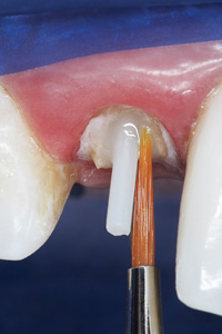
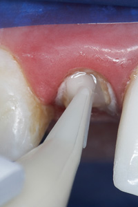
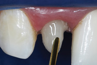
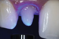
Figures 8a to 8d. (8a) A dual-curing self-etch adhesive (Futurabond DC) was applied to the remaining dentin surface and light-cured for 10 seconds. (8b) A dual-cure, radiopaque flowable core-build-up composite material (Rebilda) was injected over the coronal aspect of the post, (8c), sculpted with a long bladed interproximal instrument, (8d) and smoothed with a No. 2 sable brush to an ideal coronal preparation geometric shape and dimension and light-cured for 40 seconds.
The nonmetallic prefabricated posts have been developed as alternatives, including ceramic (white zirconium oxide) and fiber-reinforced resin posts. Zirconium oxide posts have a high flexural strength, are biocompatible, and are corrosion resistant. However, this material is difficult to cut intraorally with a diamond, and to remove from the canal for retreatment.4 The fiber-reinforced composite resin post-and-core system offers several advantages: a one appointment technique, no laboratory fees, no corrosion, negligible root fracture, no designated orifice size, increased retention resulting from surface irregularities, conserved tooth structure, and no negative effect on aesthetics.
04/01/2012 at 5:05 pm #15032Drsumitra
OfflineRegistered On: 06/10/2011Topics: 238Replies: 542Has thanked: 0 timesBeen thanked: 0 timesTHE FERRULE EFFECT
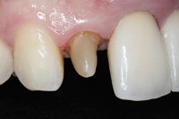
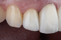
Figure 9. Completed fiber-reinforced composite post-and-core. The placement of a 1.0 mm circumferential ferrule on sound tooth structure ensures the mechanical retention and resistance.
Figure 10. An optimal adhesive integration between the components of the post-retained system which provides a structural integrity for intraradicular rehabilitation.
The successful rehabilitation of any endodontically treated tooth using the post-retained system requires the consideration of one specific structural design characteristic: the ferrule effect. The stability of the crown is influenced by the preparation design for endodontically treated teeth. Preserving tooth structure during preparation is paramount in preventing stress concentrations at the cementoenamel junction of the endodontically restored tooth and provides resistance to tooth fracture. The completed crown preparation should have a ferrule design that encapsulates the endodontically re-stored tooth complex. This collar effect provides an antirotational feature for the stability of the crown. Clinical studies have demonstrated and confirmed the importance of this coronal tooth “collar” on the mechanical resistance and retention form of the endodontically restored tooth complex.14 The general guideline is a 1.0 to 2.0 mm preparation on sound tooth structure. Procedures that provide a shoulder on tooth structure, and an axial preparation on the core buildup, will have an insufficient ferrule design. In cases where there is insufficient sound tooth structure for a ferrule design, it is necessary to obtain this dimension through periodontal crown lengthening and/or forced-tooth-eruption procedures.
CURRENT TECHNIQUES: DISCUSSION
Currently, an increased demand for clinically convenient post-and-core systems to replace lost tooth structure has provided the clinician with a plethora of simplified “one-visit” post-and-core restorative options. However, in view of the previous considerations, it is understandable that clinicians have uncertainties about selection of restorative materials and techniques to achieve optimal results for post-and-core build-up procedures.
According to studies by the CR Foundation (formerly known as Clinical Research Associates), the fiber-reinforced systems are superior to metal prefabricated posts. In the last few years, there has been a major shift away from metal custom cast post-and-cores toward resin-based composite cores.15 Prefabricated composite post systems are replacing metal post systems because an adhesive procedure with the fiber-reinforced composite post system adds strength to the tooth-restorative interface after bonding. Therefore, the fiber-reinforced post has an advantage after assembly. The fiber-reinforced composite post system has a similar modulus of elasticity to the dentin after bonding, whereas the metal post assembly has an appreciably higher modulus of elasticity. Figures 2 to 10 demonstrate the utilization of a fiber-reinforced composite post-and-core system to restore a fractured endodontically treated maxillary right central.CONCLUSION
Although the quest for the ideal material to restore lost tooth structure continues to be a focus of modern dental research, the aforementioned review indicates there are many post-and-core materials and techniques that are available to the clinician for a variety of clinical procedures and thus each clinical situation should be evaluated on an individual basis.106/01/2012 at 1:08 pm #15034 drsushant
OfflineRegistered On: 14/05/2011Topics: 253Replies: 277Has thanked: 0 timesBeen thanked: 0 times
drsushant
OfflineRegistered On: 14/05/2011Topics: 253Replies: 277Has thanked: 0 timesBeen thanked: 0 timesModified collar and buccal dentine thickness.
The effectiveness of a cervical collar with a thicker buccal dentine wall was investigated (Joseph & Ramachandran 1990). Forty extracted maxillary central incisors were divided into four groups; 1and 2 mm of buccal dentine, with and without 608 bevels. The subsequent testing w as similar to that used by Tjan & Whang (1985). The authors concluded that the use of a 2-mm collar increased the resistance of the tooth to root fracture. The authors noted there was no significant difference between the mean failure load of the two groups with cervical collars, regardless of the amount of remaining buccal dentine.Collar with resin teeth.
The effect of a cervical collar was investigated using photoelastic stress analysis on resin teeth resembling canines (Loney et al.1990). Resinwas necessary to conduct the photoelastic analysis but it must be emphasized that resin has a stiffness between a sixth to a quarter that of dentine. Eight teeth were duplicated from a mould of a master tooth. The master tooth was prepared with 2- mm incisal reduction,1.5-mm buccal reduction and an angled shoulder, and a 0.5-mm lingual chamfer. The master tooth was then reduced another 4 mm to form a flat plane for the core. Four of the resin teeth replicated this preparation; the other four had a 1.5-mm bevel around the flat plane. Cast post and cores were cemented in all the teeth. Crowns were not placed. Each tooth was then placed under a 400-gload at 1528 to its long axis, sectioned and then examined at five points. The shear stresses were less varied in the collared group, however, the teeth without collars exhibited significantly lower shear stresses at the three of the five points examined. From their findings, Loney et al. (1990) concluded that the used of a core collar did not produce a ferrule effect.Bonded posts and broken down teeth.
The influence of a metal collar on the structurally compromised teeth, with and without resin reinforcement has been examined (Saupe et al. 1996). Forty extracted maxillary central incisors were divided into two categories: resin reinforced and non-reinforced. These were further divided into roots with and without collars. The collar design was similar to that used by Barkhordar et al. (1989), having a 2-mm collar with 38 of taper on the root wall. All the teeth were prepared to simulate structurally compromised roots, with only 0.5-0.75 mm of dentine at the cementoenamel junction. The resin-reinforced roots were prepared using a visible light-cured resin composite bonded to the internal root surface. Cast post and cores were cemented in all teeth using a resin cement. All the other in vitro studies (without crowns) reviewed used zinc phosphate as a luting cement. Prior to loading the teeth, the roots were coated with rubber to simulate the periodontal ligament, then embedded in resin. The teeth were loaded until failure, which was detected by a sudden release of the load on the test tooth. Resin reinforcement significantly increased the resistance to failure, but the use of the collar in the resin reinforced group was found to be of no benefit.Torsion.
The use of a cervical collar on a post was found to be of particular benefit in increasing the resistance of the post and core to torsional forces (Hemmings et al. 1991).Where a very tapered bevel of 458 was used, an increase in resistance to torsional forces 13 times that of the control group was seen. The study did not use crowns over the cores. The authors made the important point that a metal cervical collar may be aesthetically unacceptable where the metal is visible at the gingival margin.Summary of studies without use of artificial crowns.
These studies give mixed results as to the effectiveness of a cervical collar in producing a ferrule effect (Fig.1). Even so, the results of these studies should be interpreted with caution, as none of them used artificial crowns on the cores. This was noted by Loney et al. (1990), who felt that such tests were still valid, as they helped to determine an optimal post and core form. They believed this would be of importance in crowns that had lost cement or become loose, resulting in the post and core being placed under increased forces.
Figure 1. The provision of a cervical collar as part of the core without an artificial crown. Although a beveled collar/ferrule has been depicted, a butt finish or chamfer could also be used.Despite the confusion over the usefulness of a cervical collar, cores that are made with a ferrule create technical problems. The design requires increased casting expansion at the ferruled part of the core, and a decrease in casting expansion in the post. In a comparison of casting techniques, it was demonstrated that this could not be achieved to a clinically acceptable level, despite the authors trying five different casting techniques (Campagni et al. 1993). Their results raise concern about the findings in the studies which didemploya core with a ferrule, as the technical problems encountered with casting may have influenced the outcomes of testing.
06/01/2012 at 1:11 pm #15033 drsushant
OfflineRegistered On: 14/05/2011Topics: 253Replies: 277Has thanked: 0 timesBeen thanked: 0 times
drsushant
OfflineRegistered On: 14/05/2011Topics: 253Replies: 277Has thanked: 0 timesBeen thanked: 0 timesIntroduction. Successful restoration of root-filled teeth requires an effective coronal seal, protection of the remaining tooth, restored function and acceptable aesthetics. A postretained crown may be indicated to full these requirements. However, one mode of failure of the post-restored tooth is root fracture. Therefore, the crown and post preparation design features that reduce the chance of root fracture would be advantageous. A ferrule is a metal ring or cap intended for strengthening. The word probably originates from combining the Latin for iron (ferrum) and bracelets (viriola) (Brown 1993). A dental ferrule is anencircling band of cast metal around the coronal surface of the tooth. It has been proposed that the use of a ferrule as part of the core or articial crown may be of benefit in reinforcing root¢filled teeth. A protective, or ‘ferrule effect’could occur owing to the ferrule resisting stresses such as functional lever forces, the wedging effect of tapered posts and the lateral forces exerted during the post insertion (Sorensen & Engelman1990). A literature search was conducted using the Medline database to ¢find papers that have examined the ferrule effect or made reference to it. Papers were found by searching for the word ‘ferrule’. Those pertaining to dentistry were then obtained and read to see whether they contributed in examining the ferrule effect. Some of the references used in these papers provided further articles of interest. Laboratory-based investigation of the ferrule effect. Most research investigating the ferrule effect has been conducted in the laboratory. The complexity of the oral environment prevents clear extrapolation owing to the simplicity of the experiments. Studies without use of artificial crowns. The concept of an extracoronal ‘brace’ has been proposed (Rosen 1961) and defined as a‘‘. . . subgingival collar or apron of gold which extends as far as possible beyond the gingival seat of the core and completely surrounds the perimeter of the cervical part of the tooth. It is an extension of the restored crown which, by its hugging action, prevents shattering of the root.’’ Rosen & Partida-Rivera (1996) tested this concept using7 6 extracted maxillary lateral incisors that had the crown sectioned to a level 1mm coronal to the cementoenamel junction. Half of the teeth were further prepared with a bevelled shoulder 2 mm high and 0.25 mm wide at the base, having an angle of convergence of 68. A gold casting, which represented the collar portion of a crown, was then cemented onto these teeth. Screw posts were then inserted and tightened with incremental torque until the root or post fracture occurred. The collar significantly reduced the incidence of root fracture. However, the rotational application of force in a continuous manner would be rarely present in the mouth, and implies independent movement of the post and collar. Buccal dentine thickness. The influence of dentine thickness (buccal to the post space) on the resistance to root fracture has been investigated (Tjan & Whang 1985). This study used 40 extracted maxillary central incisors divided into four groups. The control group had 1mm of remaining buccal dentine. One of the test groups also had 1mm of remaining buccal dentine and a 608 bevel. The other two test groups had 2 and 3 mm of remaining buccal dentine, and no bevel. Cast post and cores were cemented into the test teeth, but no crowns were placed. The teeth then underwent compressive loading until they failed. The incorporation of a bevel produced a core that provided a metal collar. The authors concluded from their study that the incorporation of the metal collar did not increase resistance to root fracture. No significant differences were noted between the varying dentine wall thickness, although both the groups with only 1mmof dentine all failed owing to fracture rather than cement failure. This is of particular interest as different modes of failure may be easier to manage, i.e. a loose post versus a fractured root. Modified collar. The effect of a cervical metal collar was re-examined (Barkhordar et al.1989). This study was based on that of Tjan & Whang( 1985) but used a modified collar design. Twenty extracted maxillary central incisors were divided into two groups; those with and those without a collar. Both the groups had 1mm of buccal dentine, but the test group had a 2-mm collar preparation with approximately 38 of wall taper, and a total convergence of 68. Cast post and cores were then cemented but no crowns were used. The teeth then underwent compressive loading until root fracture. Barkhordar et al. (1989) found that a metal collar significantly increased resistance to root fracture. They also observed different fracture patterns in the collared teeth compared to those without collars. The collared group predominantly underwent patterns of horizontal fracture whereas the teeth without collars mainly exhibited patterns of vertical fracture (splitting).
06/01/2012 at 1:11 pm #15035 drsushant
OfflineRegistered On: 14/05/2011Topics: 253Replies: 277Has thanked: 0 timesBeen thanked: 0 times06/01/2012 at 1:13 pm #15036
drsushant
OfflineRegistered On: 14/05/2011Topics: 253Replies: 277Has thanked: 0 timesBeen thanked: 0 times06/01/2012 at 1:13 pm #15036Anonymous
Sixty extracted maxillary incisors were used to investigate the effect of six different tooth preparation designs on the resistance to failure (Sorensen & Engelman 1990). All the teeth had post and cores cemented, over which a crown was then cemented. Each tooth was loaded at1308 to its long a fixis until it failed which encompassed either displacement of the crown or post, or fracture of the root or post.
One design was a1308 sloped shoulder from the base of the core to the margin. Whereas this design has a ferrule between the crown and margins and also between the core and tooth, it did not increase resistance to failure or fracture.
Two groups had a 90 shoulder without any coronal dentinal extension, and one of these groups also had a 1-mm-wide608bevel finish line. The placement of a bevel at the crown margin in these groups did not increase fracture resistance. This was in accordance with thefindings of Tjan &Whang (1985).
Two groups had a significantly different mean failure load compared with the other four groups. These two designs had a 908 shoulder and a 1-mm-wide 608 bevel finish line. One of the groups had a1-mm coronal dentinal extension. The other hada2-mm-wide coronal dentinal extension and a 1-mm-wide 60 contra bevel at the tooth-core junction. As failure thres hold was not significantly different between these two groups, it may be inferred that the contra bevel is of no benefit. Furthermore, and most importantly, the coronal extension of dentine above the shoulder is the design feature which increases the resistance to failure, and thus imparts a ferrule effect.
Sorensen & Engelman (1990) advised that as much coronal tooth as possible should be preserved, and a butt-joint margin between the core and tooth be used, i.e. minimal taper. They went on to suggest that the ferrule effect be defined as ‘‘ . . . a 3608 metal collar of the crown surrounding the parallel walls of the dentine extending coronal to the shoulder of the preparation. The result is an elevation in resistance form of the crown from the extension of dentinal tooth structure.’’
An investigation of root fracture related to the post selection and crown design also considered the influence of the ferrule effect (Milot & Stein1992). Forty-eight resin maxillary central incisors were divided into three groups. One of the groups had a cast post and core, the other two groups used a direct post system with a cermet cement core. Half the teeth in each group had a 1-mm concave bevel apical to the margin. Although the dimensions of the tooth preparations were not disclosed, the illustrations indicated that finall teeth there was dentine extending coronal to the margin. Therefore, the ferrule effect would have been expected in both the control and test groups in this experiment.
Crowns were cemented on all the teeth, which were then loaded with a compressive force until they fractured. The force was applied at 1208 to the long axis of each tooth. The teeth with the bevel had an increased resistance to root fracture. Milot & Stein (1992) proposed that the bevel produced a ferrule effect and the gingival extension of the metal collar provided support at the point of leverage. The use of a cermet/glass-ionomer cement as a core material, as in this study, is not recommended owing to its lack of strength (McLean 1998). The authors noted some cracking and crazing of the cement cores prior to crown placement, but did not report any significant differences in fracture resistance between the teeth with cast and cermet cement cores.Ferrule length.
Based on the effective ferrule demonstrated by Sorensen & Engelman (1990), the influence of ferrule length on resistance to preliminary failure was investigated (Libman & Nicholls 1995). The authors defined preliminary failure as the propagation of a crack in or around the luting cement of the crown. Twenty-five extracted maxillary central incisors were split into five groups; a control group and four test groups. The test groups had ferrule lengths of 0.5,1,1.5 and 2 mm. The teeth were prepared with1- mm-wide shoulders. The test teeth had cast post and cores cemented and the control group did not. All the teeth were restored with cast crowns. The teeth were subjected to cyclic loading until preliminary failure was detected, using a strain gauge. The control group and the teeth with 1.5- and 2-mm ferrules were found to be significantly better than the teeth with 0.5- and1- mm ferrules in resistance to preliminary failure. The authors concluded that1.5 mm should be the minimum ferrule length when restoring a root filled maxillary central incisor with a post- and core-retained crown.
Libman & Nicholls (1995) paid particular attention to the shortcomings of some methods of in vitro testing. The use of cyclic loading in their study was based upon the rationale that failure within the dental complex was associated with repeated fatigue loads rather than a single fracture-inducing load. The authors also acknowledged that their study did not duplicate the deformability of the periodontal ligament. Furthermore, the ferrule height in the study was constant around the circumference of the tooth, which may differ from the clinical situation where the finish line follows the morphology of the interproximal gingivae.
The conclusions of Libman & Nicholls (1995) were based on rigid parameters (Gegauff 2000). Their study used short (8-mmlong) and narrow (1.25-mmdiameter) posts and there was no simulation of periodontal support. Their finding of a1.5-mm minimum effective ferrule may not be the case clinically.
Cyclic loading was adopted in an investigation of the influence of post and ferrule length on resistance to failure (Isidor et al.1999). Using 9 0 extracted bovine teeth, they studied three post lengths (5, 7.5 and 10 mm) and two ferrule lengths (1.25 and 2.5 mm). Bovine teeth were selected in an attempt to reduce variability. The teeth were restored with prefabricated titanium posts and resin composite cores. The teeth were all crowned, then subjected to cyclic loading until the crown or post dislodged or the post or root fractured. The fracture resistance of the specimens increased with the ferrule length, but was not enhanced by increased post length. The authors note that this is of particular significance in re-evaluating studies which have investigated post length and design without a crown in their experiment methodology. Furthermore, it would suggest that an increase in post length, to achieve an increase in retention does not decrease resistance to failure.Type of post and core.
Most of the studies investigating the ferrule effect have used cast post and cores. Milot & Stein (1992) used direct posts with cermet cores, but found no significant difference in fracture resistance compared with the teeth they restored with cast post and cores. The posts were all cemented with zinc phosphate cement. Al-Hazaimeh & Gutteridge (2001) investigated the effect of a ferrule on central incisors when a direct post and composite resin core was used. The prefabricated posts and composite resin cores were placed in 20 central incisor teeth. The posts and crowns were luted with a resin cement. Ten of the teeth had a 2-mm ferrule, the others had no ferrule. Analysis following compressive loading until failure demonstrated no significant difference between the two groups. The authors proposed that the strength afforded by the resin may have masked any benefit provided by the ferrule.
The authors observed a high mean failure load for both groups, which they attributed to the resin luting cement. Although there was no difference between the two groups’resistance to failure, the modes of failure differed. The group with a ferrule underwent oblique fracture, whereas the group without a ferrule largely underwent vertical root fracture.06/01/2012 at 1:14 pm #15037Anonymous
Summary of studies with artificial crowns.
These studies showed that coronal extension of dentine above the shoulder increases the resistance to failure (Fig.2).This ferrule effect may be significantly enhanced when the coronal extension of dentine is at least 1.5 mm. This effect has been demonstrated in teeth restored with cast post and cores. A ferrule in teeth restored with direct posts and resin cores may not offer any additional benefit.
In all the in vitro studies, both with and without crowns, only single rooted teeth have been investigated. The influence of the ferrule effect on multi-rooted teeth remains an area for further research.The ferrule effect in vivo.
There appear to be no reports of prospective clinical investigations of the ferrule effect. A retrospective study of the survival rate of two post designs was conducted by Torbjorner et al. (1995).Their study involved reviewing the records of 638 patients, with a total of 788 posts; the patients were not examined. Seventy-two of the posts failed. Most (46 cases) failed owing to the loss of retention. It was observed that all the post fractures (six cases) occurred in teeth with a ‘‘.. lack of a ferrule effect of the metal collar at the crown margin area…’’. The remainder failed owing to root fracture. Their study did not state how many crowns surveyed had a ferrule. From the work of Sorensen & Engelman (1990) and Libman & Nicholls (1995) it has been shown that the position and length of the ferrule is of significance. Torbjorner et al. (1995) did not indicate whether these design features were recorded in the patients’ files. Furthermore, radiographs do not allow assessment of the amount of tissue remaining under a crown with a metal substructure. Therefore, it would be prudent for future studies to make some record of the ferrule, oreven consider keeping dies.
A classification of the single-rooted pulpless teeth based upon the amount of remaining supra gingival tooth structure has been recommended (Kurer 1991). Five classes of pulpless tooth were described; class I has sufficient coronal tissue for a crown preparation, II has coronal tissue but requires a core, and III has no coronal tissue. The classes IV and V refer to the complications of intra osseous fracture and periodontal disease. However, this classification does not account for a minimum effective ferrule length. It would perhaps be of value to add a further subclassification of ferrule length to the Class II type of pulpless tooth. A suitable distinction would be less than or at least2 mm-ferrule length.Preoperative tooth assessment.
In assessing whether a tooth should be restored, the clinician must consider the amount of remaining supragingival tissue. Libman & Nicholls (1995) demonstrated in vitro that the minimum effective ferrule should be 1.5 mm of coronal dentine extending beyond the preparation margin. Below this length, there is a significant decrease in resistance to preliminary failure. Without supportive in vivo research the clinician is left to question whether satisfactory treatment can still be provided where a ferrule is absent or that is shorter than that advised in this in vitro study.A minimum ferrule length of 2 mm to compensate for the difficulties of intraoral tooth preparation has been recommended (McLean1998). Citing a paper by Freeman et al. (1998), it was noted that even where a1-mmferrule was attempted extraorally, it was difficult to achieve. Overcompensation is thus advisable to achieve an adequate length of parallel dentine, to produce an effective ferrule.
The ferrule length that may be obtained will be influenced by the ‘biologic width’. This is defined as ‘‘. . . the dimension of the junctional epithelial and connective tissue attachment to the root above the alveolar crest’’ (Sivers & Johnson 1985). If unpredictable bone loss and inflammation is to be avoided, the crown margin should be at least 2 mm from the alveolar crest. It has been recommended that at least 3 mm should be left to avoid impingement on the coronal attachment of the periodontal connective tissue (Fugazzotto & Parma-Benfenait 1984).Therefore, at least 4.5 mm of supra-alveolar tooth structure may be required to provide an effective ferrule.
In those clinical situations where there is insufficient ferrule length, even where margins are placed subgingivally, the clinician may consider surgical crown lengthening or orthodontic extrusion. This allows the distance between the crown margin and alveolar crest to bewidened, and increases the potential ferrule length. Methods to increase ferrule length will reduce the root length and result in more tooth loss, possibly making the crown to root ratio unfavourable. Furthermore, both procedures will add to the cost of restoring the tooth, prolong treatment time and cause discomfort to the patient. The study by Al-Hazaimeh & Gutteridge (2001) suggests that the use of a resin-bonded direct post and resin core may be a preferable alternative where a ferrule can not easily be obtained.
The effect of crown lengthening to establish a ferrule on static load failure was investigated (Gegauff 2000). Using restorative composite resin to make root analogues, two groups of10 teeth were tested. One group simulated teeth that had received crown lengthening and a ferrule in the preparation; the other group were not crown lengthened and were without a ferrule and no remaining clinical crown. All the teeth had cast post retained cores and crowns cemented prior to testing. Both the alveolar bone and periodontal ligament were simulated in the study. The teeth were subjected to a load by a universal testing machine until failure (peak load). The group that had crown lengthening and a ferrule demonstrated a significantly lower failure load.
Gegauff (2000) noted that the apical relocation of the finish line following crown lengthening resulted in a decrease in the cross section of the preparation. Subsequently, this reduction in tissue combined with an altered crown to root ratio may result in weakening of the tooth. He also pointed out that orthodontic extrusion may be preferable to crown lengthening as it results in a smaller change in the crown to root ratio. The results of that study suggested that the overall importance of a ferrule at the expense of remaining tooth structure was unclear.
A balance between the ferrule length obtained and the remaining root is there for eneeded. These considerations are obviously best made prior to root canal treatment. If a suitable ferrule length cannot be obtained, the patient should be informed of the potential compromise.06/01/2012 at 1:15 pm #15038 -
AuthorPosts
- You must be logged in to reply to this topic.