Home › Forums › Oral Diagnosis & Medicine › TREATMENT FOR LICHEN PLANUS
Welcome Dear Guest
To create a new topic please register on the forums. For help contact : discussdentistry@hotmail.com
- This topic has 3 replies, 1 voice, and was last updated 23/04/2012 at 5:23 pm by
Drsumitra.
-
AuthorPosts
-
23/04/2012 at 5:15 pm #10464
Drsumitra
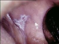
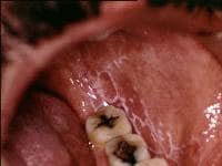
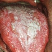
OfflineRegistered On: 06/10/2011Topics: 238Replies: 542Has thanked: 0 timesBeen thanked: 0 timesMedical treatment of oral lichen planus (OLP) is essential for the management of painful, erythematous, erosive, or bullous lesions. The principal aims of current oral lichen planus therapy are the resolution of painful symptoms, the resolution of oral mucosal lesions, the reduction of the risk of oral cancer, and the maintenance of good oral hygiene. In patients with recurrent painful disease, another goal is the prolongation of their symptom-free intervals.[30, 31, 32]
The main concerns with the current therapies are the local and systemic adverse effects and lesion recurrence after treatment is withdrawn. No treatment of oral lichen planus is curative.
Eliminate local exacerbating factors. Treat any sharp teeth or broken restorations or prostheses that are likely to cause physical trauma to areas of erythema or erosion by using conventional dental means. Scale the teeth to remove calculous deposits and reduce sharp edges. If the patient has an isolated plaquelike or erosive oral lichen planus lesion on the buccal or labial mucosa adjacent to a dental restoration, and if an allergy is detected by means of skin patch testing, the lesion may heal if the offending material is removed or replaced. (However, most lichenoid lesions adjacent to dental restorations are asymptomatic.)
If systemic drug therapy (eg, treatment with NSAIDs, antimalarials, or beta-blockers) is suspected as the cause of oral lichenoid lesions, changing to another drug may be worthwhile. This change must be undertaken only by the patient’s attending physician. However, the switch rarely resolves the erosions, and almost never resolves the white patches of oral lichen planus.
Inform all patients with oral lichen planus about their slightly increased risk of oral SCC (the most common of all oral malignancies). As with all patients, advise those with oral lichen planus that this risk may be reduced by eliminating tobacco and alcohol consumption and by consuming a diet rich in fresh fruits and vegetables, among other measures (see Complications). Erosive and atrophic lesions can be converted into reticular lesions by using topical steroids. Therefore, the elimination of mucosal erythema and ulceration, with a residual asymptomatic reticular or papular lesions, may be considered an end point of current oral lichen planus therapy. With respect to plaque lesions, the effect of treatment on the risk of oral cancer is unclear.
Topical corticosteroids are the mainstay of medical treatment of oral lichen planus, although rarely, corticosteroids may be administered intralesionally or systemically. Some topical corticosteroid therapies may predispose the patient to oral pseudomembranous candidosis. However, this condition is rarely if ever symptomatic, and it generally does not complicate healing of the erosions related to oral lichen planus. Topical antimycotics (eg, nystatin, amphotericin) may be prescribed when an infection is present.Erosive oral lichen planus that is recalcitrant to topical corticosteroids may respond to topical tacrolimus. Other potential therapies for recalcitrant oral lichen planus include hydroxychloroquine, azathioprine, mycophenolate, dapsone, systemic corticosteroids, and topical and systemic retinoids.
23/04/2012 at 5:21 pm #15420Drsumitra
OfflineRegistered On: 06/10/2011Topics: 238Replies: 542Has thanked: 0 timesBeen thanked: 0 times23/04/2012 at 5:22 pm #15421Drsumitra
OfflineRegistered On: 06/10/2011Topics: 238Replies: 542Has thanked: 0 timesBeen thanked: 0 timesCurrent data suggest that oral lichen planus is a T-cell–mediated autoimmune disease in which autocytotoxic CD8+ T cells trigger apoptosis of oral epithelial cells.[1, 2]
The dense sub-epithelial mononuclear infiltrate in oral lichen planus is composed of T cells and macrophages, and there are increased numbers of intra-epithelial T cells. Most T cells in the epithelium and adjacent to the damaged basal keratinocytes are activated CD8+ lymphocytes. Therefore, early in the formation of oral lichen planus lesions, CD8+ T cells may recognize an antigen associated with the major histocompatibility complex (MHC) class I on keratinocytes. After antigen recognition and activation, CD8+ cytotoxic T cells may trigger keratinocyte apoptosis. Activated CD8+ T cells (and possibly keratinocytes) may release cytokines that attract additional lymphocytes into the developing lesion.[2]
Oral lichen planus lesions contain increased levels of the cytokine tumor necrosis factor (TNF)–alpha.[3, 4] Basal keratinocytes and T cells in the subepithelial infiltrate express TNF in situ.[5, 6] Keratinocytes and lymphocytes in cutaneous lichen planus express elevated levels of the p55 TNF receptor, TNF-RI.[7] T cells in oral lichen planus contain mRNA for TNF and secrete TNF in vitro.[8] Serum and salivary TNF levels are elevated in oral lichen planus patients.[9, 10, 11, 12] TNF polymorphisms have been identified in patients with oral lichen planus, and they may contribute to the development of additional cutaneous lesions.[13] Oral lichen planus has been treated successfully with thalidomide,[14, 15] , while thalidomide is known to suppress TNF production.[16, 17] Together, these data implicate TNF in the pathogenesis of oral lichen planus.
The lichen planus antigen is unknown, although it may be a self-peptide (or altered self-peptide), in which case lichen planus would be a true autoimmune disease. The role of autoimmunity in the pathogenesis is supported by many autoimmune features of oral lichen planus, including its chronicity, onset in adults, predilection for females, association with other autoimmune diseases, occasional tissue-type associations, depressed immune suppressor activity in patients with oral lichen planus, and the presence of autocytotoxic T-cell clones in lichen planus lesions. The expression or unmasking of the lichen planus antigen may be induced by drugs (lichenoid drug reaction), contact allergens in dental restorative materials or toothpastes (contact hypersensitivity reaction), mechanical trauma (Koebner phenomenon), viral infection, or other unidentified agents.
23/04/2012 at 5:23 pm #15422Drsumitra
OfflineRegistered On: 06/10/2011Topics: 238Replies: 542Has thanked: 0 timesBeen thanked: 0 timesRe-examine patients with oral lichen planus (OLP) during active treatment, and monitor lesions for reduction in mucosal erythema and ulceration and alleviation of symptoms. Continue active treatment and try alternative therapies until erythema, ulceration, and symptoms are controlled. Follow up with patients with oral lichen planus at least every 6 months.
Advise patients with oral lichen planus to pay attention to when symptoms are exacerbated or when lesions change. Such changes generally indicate a phase of increased erythematous or erosive disease.
In view of the potential association of oral lichen planus with oral SCC, an appropriate specialist should follow up with the patients every 6-12 months. In addition, advise patients to regularly examine their mouths and seek the help of a specialist if persistent red or ulcerative oral mucosal lesions develop.
Candidal cultures or smears may be obtained periodically. Infections can be controlled with topical antimycotic preparations. These tests may be of limited clinical value because oral C albicans is present in at least 70% of all healthy persons.
-
AuthorPosts
- You must be logged in to reply to this topic.


